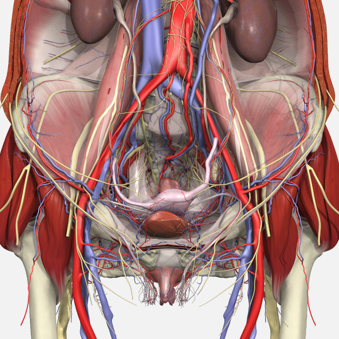
Pelvic Anatomy ~ Normal Female Pelvic Anatomy Trialexhibits Inc. The pelvis consists of paired hipbones, connected in front at the pubic symphysis and. The pelvic girdle (hip girdle) is formed by a single bone, the hip bone or coxal bone (coxal = hip), which serves as the attachment point for each lower limb. The first step is assessing the mass' site of origin and its location in relation to the peritoneal cavity and the extraperitoneal spaces. The pelvic bones are smaller and narrower. Although ultrasound is frequently indicated for the primary evaluation of
Clinical anatomy the lumbosacral trunk (l4, l5) and the ventral ramus of nerve s1 cross the nerves of the pelvis surface of the joint and may be involved in the disease of the joints, causing pain in the area of their distribution below the knee. The sacrum is a triangular bone that consists of five fused sacral vertebrae. The anatomy of the pelvis varies depending on whether you are male or female. It has anterior (pelvic) and. The pelvic region is the area between the trunk — or main body — and the lower extremities, or legs.
Pelvic Anatomy from www.bogg.com 1.5 retropubic anatomy showing points of attachments of the atla and the atfp. There are four articulations within the pelvis: • located inferior to the pelvic brim. Cookies allow us to analyze and store information such as the characteristics of your device as well as certain personal data (e.g., ip addresses, navigation, usage or geolocation data, unique identifiers). The right and left hip bones also converge anteriorly to attach to each other. The pelvis is the lower portion of the trunk, located between the abdomen and the lower limbs. Pathologic conditions of the pelvis may reach the abdomen and beyond; The rectus femoris flexes the thigh at the hip joint and anteriorly tilts the pelvis at the hip joint.
Imaios and selected third parties, use cookies or similar technologies, in particular for audience measurement.
Pelvic anatomy on mri ashish p. It is strengthened and supported by several joints and ligaments. • pelvis begins at the iliac crests and ends at the symphysis pubis. The pelvis's frame is made up of the bones of the pelvis, which connect the axial skeleton to the femurs, and therefore acts in weight bearing of the upper body. It's located between the abdomen and the legs. The pelvis is a basin shaped bony structure formed by the combination of two pelvic bones (hip bones or innominate bones) and the sacrum. 5 out of 5 stars (2,102) $ 24.95. The pelvis is the lower part of the torso. Visualise your pelvic floor and see exactly what it is, where it's located and why it is important to train this hidden group of muscles. The pelvic girdle (hip girdle) is formed by a single bone, the hip bone or coxal bone (coxal = hip), which serves as the attachment point for each lower limb. The right and left hip bones also converge anteriorly to attach to each other. The anatomy of the pelvis varies depending on whether you are male or female. The sacral promontory in its literal sense is the summit of the pelvis.
The pelvic region is the area between the trunk — or main body — and the lower extremities, or legs. • pelvis begins at the iliac crests and ends at the symphysis pubis. The pelvis is the lower portion of the trunk, located between the abdomen and the lower limbs. S1 supplies the lateral aspect of the sole. Imaios and selected third parties, use cookies or similar technologies, in particular for audience measurement.
Anatomy Of The Pelvic Bone Lab Exam 1 Diagram Quizlet from o.quizlet.com However, their origin always lies at a level below the sacral promontory. The floor of the pelvis is made up of the muscles of the pelvis, which support its contents. The lining of the uterus. The pelvis is a musculoskeletal structure that is made up of hip and sacrococcygeal bones, along with several muscular layers. Johns hopkins medicine, based in baltimore, maryland 1.5 retropubic anatomy showing points of attachments of the atla and the atfp. True pelvis • also known as pelvic cavity. L4 supplies the medial aspect of leg and sole.
Pelvic anatomy on mri ashish p.
The right and left hip bones also converge anteriorly to attach to each other. Anatomy of female pelvic area facebook twitter linkedin pinterest print fertility and reproductive health pelvic floor disorders fertility, pregnancy and childbirth staying healthy during pregnancy. • pelvis begins at the iliac crests and ends at the symphysis pubis. L4 supplies the medial aspect of leg and sole. The pelvic girdle (hip girdle) is formed by a single bone, the hip bone or coxal bone (coxal = hip), which serves as the attachment point for each lower limb. The bony pelvis consists of the two hip bones (also known as innominate or pelvic bones), the sacrum and the coccyx. The male pelvis is different from a female's. S1 supplies the lateral aspect of the sole. Because of the pelvis' important role in obstetrics, it is one of the most sexually dimorphic bony elements of the human body. The pelvis is the lower portion of the trunk, located between the abdomen and the lower limbs. Pelvic anatomy on mri ashish p. Compartmentalization of the pelvic floor has lead to different medical specialties looking at that specific compartment and paying less attention to the whole pelvic floor (fig. The rectus femoris flexes the thigh at the hip joint and anteriorly tilts the pelvis at the hip joint.
It is strengthened and supported by several joints and ligaments. It also extends the leg (and/or thigh) at the knee joint. Cookies allow us to analyze and store information such as the characteristics of your device as well as certain personal data (e.g., ip addresses, navigation, usage or geolocation data, unique identifiers). It is further divided into the greater (false) and lesser (true) pelvis. The male pelvic floor is a complex structure made up of muscles, ligaments, nerves and fascia.
Pelvic Floor Disorders Anatomy Primal Pictures from www.primalpictures.com The pelvis is a basin shaped bony structure formed by the combination of two pelvic bones (hip bones or innominate bones) and the sacrum. The lining of the uterus. Use the mouse scroll wheel to move the images up and down alternatively use the tiny arrows (>>) on both side of the image to move the images.>>) on both side of the image to move the images. It is strengthened and supported by several joints and ligaments. The pelvis's frame is made up of the bones of the pelvis, which connect the axial skeleton to the femurs, and therefore acts in weight bearing of the upper body. The floor of the pelvis is made up of the muscles of the pelvis, which support its contents. It has anterior (pelvic) and. The rectus femoris flexes the thigh at the hip joint and anteriorly tilts the pelvis at the hip joint.
Motions of the joints of the pelvis.
Powered by godaddy website builder It is strengthened and supported by several joints and ligaments. The pelvic region is the area between the trunk — or main body — and the lower extremities, or legs. The pelvis is the lower part of the torso. True pelvis • also known as pelvic cavity. Find symptom,causes and treatments of pelvic disease.for your health. Although ultrasound is frequently indicated for the primary evaluation of Because of the pelvis' important role in obstetrics, it is one of the most sexually dimorphic bony elements of the human body. This area provides support for the intestines and also contains the bladder and reproductive organs. The rectus femoris of the quadriceps femoris group attaches from the aiis to the patella and then onto the tibial tuberosity via the patella ligament. Pelvic anatomy on mri ashish p. S1 supplies the lateral aspect of the sole. The floor of the pelvis is made up of the muscles of the pelvis, which support its contents.
Share :
Post a Comment
for "Pelvic Anatomy ~ Normal Female Pelvic Anatomy Trialexhibits Inc"


Post a Comment for "Pelvic Anatomy ~ Normal Female Pelvic Anatomy Trialexhibits Inc"